ESSAY-1
INTRODUCTION
Cancer is known as fatal diseases in the world , it cause more the twenty five percent of death in united kingdom. These number are increasing continuously due to prolonged life expectancy, population growth and more number of risk factors as like lack of activity, more number of obesity, and smoking (Murugan, 2019). A general characteristic of most tumours is a very few level of oxygen, is known as hypoxia, the seriousness of which depends upon the kinds of tumour. In expanding tumour or intensively proliferating tissue, demand of oxygen is more than the supply of oxygen, the existing vasculature and the distance between the cells are also increases. Therefore, it creates obstruction in the diffusion of oxygen and making even more hypoxic condition. In the hypoxic tumour cells poorer oxygen condition than the normal cells which is generally 1-2% O2 and below (Du, and et. al., 2021). Oxygen level of tumour is depends upon the stage, size, method of oxygen measurement, initial oxygenation of the cells and the measurement which was performed. This essay will going to discuss about the role of hypoxia in the regulation of angiogenesis in cancer. This essay will also include the key factors in the process of regulation of angiogenesis because of hypoxia in cancer cell. Finally this essay will argue about those pathway which is involved in the process of regulation of angiogenesis due to hypoxia in cancerous cell (Ma, and et. al., 2020).
MAIN BODY
Generally, hypoxia is a condition in cancerous cell, in which oxygen supply to cancerous cell is less than the demand of oxygen. A dissertation outline generator can help organize research on how cancerous cells commonly respond in various ways towards lowering oxygen level leading to cell survival or cell death, it is partially depends on the exposure time to hypoxia. Acute hypoxia is short term hypoxia and it occurs when occlusion of blood vessel lasts for several minutes at least. In acute hypoxia, cells have been allowed to survive in adverse situation through activating autophagy, which is a metabolic and apoptotic adaptation of cells (Mortezaee, 2020). It is gained by lowering the oxidative metabolism. On the other hand cycling hypoxia led to enhanced ROS (reactive oxygen species) generation, which contribute to the progression and survival of tumour cell. Both long term and short term hypoxia are associated with an increase in radio-resistance of cancerous cell. Acute hypoxia is related with a more aggressive phenotype of tumour (Joseph, and et. al., 2018). In the earlier stage of malignant progression, a tumour is placed in the inactive stage in a non-vascular space at which the pace of cell differentiation is balanced by the cell apoptosis. Later the tumour cells undergoes a change to form a non-angiogenic stage to active stage and this process happens through a so-called angiogenic switch (Chang, and et. al., 2020). For the progression of tumour cells, angiogenesis is important because the growth of the tumour cells often outstrips the supply of nutrients and oxygen. Angiogenesis plays a vital role in both the formation and progression of tumour. Tumour vessels are more leaky, poorly functioning, and highly irregular, despite active angiogenesis. Even in more vascular tissue, this leads to HIF-a stabilisation and hypoxic domains, even in more vascular tissue (Zhu, and Zhang, 2018). HIF activation and hypoxia have intense effects on the biology of tumour, because the expression of HIF-1Aa or HIF-2a is associated with metastatic illness and less prognosis in a number of cancers, involving colon, liver, neck and head, skin, brain, pancreas, and breast. This is due to partially therapeutic resistance because it is stubborn to treat the hypoxic tumour through radiation therapy as molecular oxygen is needed for the cytotoxic effects of radiation therapy (Kochan-Jamrozy, and et. al., 2019). Due to the HIF-mediated expression of ATP-dependent efflux pump of drugs and poor delivery of drugs, chemotherapy is also not effective in hypoxic tumours. HIF pathway and Hypoxia activation in cancerous tissues are a vital stimulus for the growth of blood vessels. HIF-1a and HIF-2a control the expression of a suite of pro-angiogenic genes, including Ang-1, Ang-2, Vegf, and Tie-2, numbers of which themselves work as biomarkers for tumour hypoxia. Ang-1 boosts tumour angiogenesis by engaging pericytes for the maturing vessels. Extra expressed in glioma, breast cancer, and stem cell factor is a HIF-1 alpha transcriptional target which arbitrates neovascularization by increasing EC migration and survival and mobilization of EPC (Klaunig, 2018). As well as the well-defined pro-angiogenic genes controlled by the HIF. According to a recent study, additional effector molecules can be regulated by hypoxia, which maximises the regulatory capacity of HIF (Sahebi, and et. al., 2020). In the growth of angiogenesis in cancer, some growth factors are involved such as; acidic & basic fibroblast growth factor, vascular endothelial growth factor family, angiostatin, angiogenin, transforming growth factor, tumour necrosis factor-alpha, and so on. Platelet-delivered endothelial growth factor, hepatocyte growth factor, epidermal growth factor, placental growth factor, and granulocyte colony-stimulating factors also play a vital role in the regulation of angiogenesis in cancer cells. Some factors work as inhibitors for tumour cells like cytokines, proteases, and protease inhibitors (Kim, and et. al., 2018). There are various proteins which occur naturally and can inhibit angiogenesis, involving endostatin, interferon, platelet factor 4, angiostatin, prolactin 16kd fragment, thrombospondin, and metalloproteinase-1, -2, & -3 tissue inhibitors. By one or more fragments of plasminogen, Angiostatin is formed. It promotes apoptosis in tumour cells and endothelial cells, and stops the formation and migration of tubules in endothelial cells.
 |
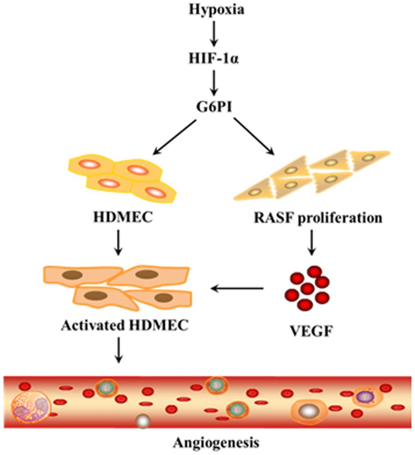
Illustration 1: steps of hypoxia and role of G6P1
Angitensin treated tumours' immunohistochemical examination point out a lowering in the expression of mRNA for bFGF and VEGF (Devarajan, Manjunathan, and Ganesan, 2021). Enostatin, it is a twenty kilodalton C-terminal fragment of collagen of type XVII, it is a part of the basement membrane. It induce the migration and proliferation of endothelial cells and also inhibits the growth factors like bFGF and VEGF-A. Since there are two types of factors which affects the angiogenesis growth that one is angiogenesis promoter and another one is angiogenesis inhibitors. When the balance between these two factor are disturb in the body then either it provokes or inhibits the process of angiogenesis in tumour cells (Luo, and Wang, 2019). When promoter increased then enhances the growth of tumour cells but on increasing the number of inhibitors, it inhibits the growth of tumour cells. VEGF-A, that is a Vascular endothelial growth factor A is a main regulator of angiogenesis in disease condition and under normal both. It belongs to a gene factors' family which also surrounds VEGF-B, VEGF-C, VEGF-D, VEGF-E and PIGF ( placenta growth factor). All of these factors have various level of specificity and different affinities for VEGFR that is tyro-kinase receptors -1,-2,&-3. when the binding of VEGF-A with VEGFR-2 then it leads angiogenesis (Zhu, and Zhang, 2018). In addition, VEGF-C and VEGF-D prefer to bind with VEGFR-3, which expressed more dominantly on lymphatic EC, that results in the differentiation of lymphatic vessels (Sahebi, and et. al., 2020). VEGF play a role in exceeding the angiogenesis in cancer by a complex paracrine and autocrine signalling pathway. VEGF show a vital role in promoting the functions of stem cell of cancer and also in the initiation of tumour. In hypoxic tumour, TAMs (tumour associated macrophages) secrete VGEF and known for its protumour functions (Minassian, and et. al., 2019). In the tumour micro environment, VEGF interacts with essential immune cells which is regulatory T cells and CD4+ forkhead box protein p3 that is FOXP3, it is a very good suppressor of immunity for anticancer. In TME, more number of fibroblasts are present and known for support growth of tumour, also release VEGF (Schito, 2019).
 |
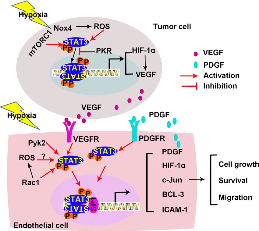
Illustration 2: steps involves in hypoxia induce angiogenesis in cancer
Source: https://www.researchgate.net/figure/STAT3-in-hypoxia-induced-angiogenesis-Hypoxia-activates-STAT3-in-both-tumor-cells-and_fig4_318914913
CONCLUSION
According to the above discussion it has been concluded that hypoxia is a condition where demand of the oxygen is more then the supply of oxygen. Exposure time of cancerous cell to hypoxia play a vital role in terms of deciding the fate of that cells that is leading to death of cells or survival of cells. Hypoxia lead to generation of reactive oxygen species cells which decide the survival and death of cancerous cells. In this essay, It has been seen that in terms of cancerous cell progression hypoxia played a very crucial role. When the balance between cancerous cell promoter and cancerous cell inhibitor disturb in the human body then it can cause angiogenesis in cancer on increasing the number of cancerous cell. But when the number of inhibitor increases then it suppress the activity of cancerous cell. VEGF is a type of promoter of cancerous cell where and interferon and many more like this are inhibitors for cancerous cell growth. In this essay the role of hypoxia in the progression of angiogenesis in cancer has been discussed with the key factor involved in the regulation of angiogenesis in cancer. Different pathways responsible for angiogenesis in cancer has also been discussed in this essay.
ESSAY-2
INTRODUCTION
Metastasis, it is one of the major reasons of death worldwide. At the time of development of tumour, cancer cells come into genetic mutations, adopt their micro-environment and promote angiogenic germination which can effectively lead to metastasis. Solid tumour's metastatic progression can be split into 5 main steps, which are: (1) Invasion of the cell migration and basement membrane, (2) Intravasation into the neighbouring lymphatic or vasculature system, (3) Staying alive in the lymphatic or vasculature circulation, (4) Discharge from vascular system to secondary tissue and in last (5) formation of colony at the site of secondary tumour (Yu, and et. al., 2019). Every stages of metastasis forces various, frequently worst conditions potentially taxing challenging to the cancerous cell to complete (Cortés-Hernández, Eslami-S, and Alix-Panabières, 2020). The number of live cancerous cell which survive and well complete all stage lowering precipitously, as the cascade progresses. In this essay several molecular event involved at various steps in the metastatic cascade will be include. Different way to treat the cancer cell through the assist of the molecular events includes in the metastatic cascade will going to describe in this essay.
Related Samples: Occupational Hygiene, Health and Ergonomics
MAIN BODY
The metastatic cascade explains as the procedure by which aggressive cancerous cell leave the primary tumour, by travelling via the bloodstream, and at the end reach the distant body parts to evolve one or more metastases. There are several steps include in the metastasis of cancer. These steps involves separation from the primary tumour, invasion via basements membranes or neighbouring tissues, entry and live in the vascular circulation, peritoneal space or lymphatics and arrest in the distant end target body parts (Berish, and et. al., 2018). Step of metastatic cascade and molecular events involved for cancer treatments are following: Step 1: invasion and migration: Metastasis is started at the time of invasion and migration where cancer cells puncture the basement membrane and cross as single cells or through collective that is via stromal micro-environment. Invasion by the basement membrane is known as differentiating steps to separate precancerous neoplasia & malignant tumour in which enhance fibre thickness, linearized fibre architecture and collagen deposition hand out to a stiffer condition (Lu, and et. al., 2019). By the cycle of contraction and protrusion cell mechanically remodel ECM, and by using metalloproteinases cells chemically deteriorate the matrix as they migrate. Apart from this, matrix stiffness and cancer cell contractility made a positive feedback loop resulting downstream impacts on behaviour of cell at the time of progression of metastatic. Through the use of organoids partially reduce these restriction through more better representing phenotypic and genotypic diversity in structure. Organoids seizes number of genomics variations present in the solid tumours and act as preclinical drug screening cancer model, shown to establish a relationship with clinical response to common therapeutics of cancer. In additionally, spheroids are the groups of cells which used to study invasion and migration (Garner, and de Visser, 2020). Stability and reproducibility make these appropriate to identifying characteristic of the cancer micro-environment which derive metastatic behaviour of cell.
 |
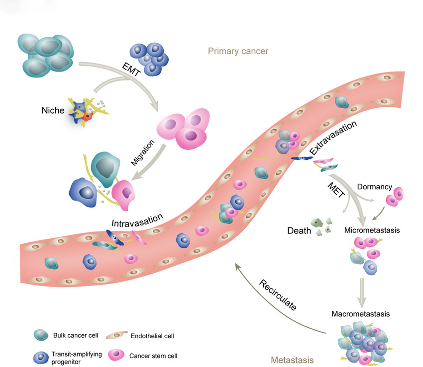
Illustration 3: Metastatic cascade
Source: https://www.google.com/search?q=metastatic+cascade+in+cancer&tbm=isch&ved=2ahUKEwjYvoS4lYf5AhWfi9gFHSehCegQ2-cCegQIABAA&oq=metastatic+cascade+in+cancer&gs
Induction of hypoxia in a spheroid model was seen to be necessary in obtaining the tumour stem like cell phenotype, a main target in present therapeutics of cancer (Zhang, and et. al., 2019). Through modelling the relationship in between the cancer micro-environment and the cell behaviour, number of therapeutics can be evolved. Spheroid model can also give a platform to study the differentiating elements between collective and single cell migration. Myoepithelial cells neighbouring the basement membrane are thought to keeps a suppressor of tumour role which can be lost at the time of neoplasia. Tumour cells can put in directly into collagen bed to evaluate the direction, morphology and cell speed at the time of migration. Significantly there is now important evidence to put forward that collagen matrix alignment is a autograph of metastatic cancer and can be utilise to forecast result of patient (Berish, and et. al., 2018). Step 2: Angiogenesis and intravasation: Angiogenesis of tumour touch on the origination of vasculature at the time of progression of tumour, allowing delivery of oxygen and nutrients and as well as excretion of the waste product. Due to absent of basement membrane and perivascular coverage, newly formed cancer vasculature is more permeable and less mature resulting leakage of proteins present in plasma which is further clear the way intravasation of tumour cell and formation of new vessel. Angiogenesis promote metastasis through empowering transport of cancer cells to distant site through lymph and vascular systems. As like perfusable models which empower the production of endothelial networks application in the recognition of the specific influences of angiogenesis on the progression of cancer. In lab assays of angiogenesis concentrate first and foremost on cell migration, endothelial barrier integrity, cell proliferation and vessel formation (Li, and et. al., 2019). In the recent time more advanced models have been evolved to learn barrier function and patient specific formation of endothelial tubule by endothelial embedding cells, within three dimensional hydrogels, including sample of patient derived. Because of interstitial pressure and blood flow promote cancer angiogenesis, number of micro-fluidic systems concentrates on recapitulating this in vitro (Riggi, Aguet, and Stamenkovic, 2018). In addition very large number of inter-tumour heterogeneity in the activity of angiogenic depends partially on those organ from where it arose and subtype of cancer, due to difference of organ specific in the anti and pro angiogenic molecule secretion outline of stromal cell populations. Therefore, the development of very unique individualized experimental system will empower characterization of angiogenic habits in various kinds of cancer for single patients, leading to enhancements in the models of drug screening. Micro-fluidic system gives permission to the amalgamation of fluid flow and are compliant to current time imaging capability (Nishida, and et. al., 2020). Step3: live in the circulation and attachment to the endothelium: However, some number of cancerous cells reaches in the body fluid circulation, even very less number of these are survive the immune stress, red blood cell collisions and haemodynamic shear forces they come up against once there. CTC (circulating tumour cells) are arrest in a circulatory vessels and extravasate by two primary mechanisms: adhesion after rolling and physical occlusion. At the time of physical occlusion, a circulating tumour cells' diameter exceed that of the micro vasculature, and the cell became lodge at the time before extravasating and attaching (Dongre, and Weinberg, 2019). At the time of rolling- adhesion, circulatory tumour cells collide to the endothelium, roll through P-selectin or E- selectin binding, and arrest through vascular cell adhesion molecule-1 (VCAM-1) or by intercellular adhesion molecule-1 ( ICAM-1) binding (Steinbichler, and et. al., 2020). Micro tubing and micro fluidic systems enabling the gathering of both clustered and single circulatory tumour cells from the blood of patient have contributed effectively to understanding the metastasis of cancer. These stage frequently take on surfaces functionalized with the circulatory tumour cells specific adhesion antibodies and proteins to improve adhesion dynamics for circulatory tumour cells at the time of reducing that of leukocytes also associated in the whole blood (Li, and et. al., 2020). Some of the physical entrapment in the flow may also be use to separate the clusters of circulatory tumour cells which have been suggested to have enhanced metastatic capability as compare to single circulatory tumour cells. A three dimensional printing of carbohydrate glass sacrificial fibre may make more perfusable, controlled and controlled vascular networks . Live cell lithography technique was evolved for better control of tumour cells. By the correlation of clinical data, theranostic stages with circulatory tumour cells seperated from patient blood have the capability to enhance the clinical results. (4) Extravasation and colonization: Go along with arrest in the circulatory vessel, tumour cells must extravasate to the colonize new site from the circulatory vessel (Wee, and et. al., 2019). This process not similar to the intravasation, in which tumour cells navigate tumour modified stroma through durotactic and chemotactic gradients in the direction of leaky, nascent vasculature with not feeling haemodynamic stressors rather the vasculature during extravasation which is broke by cancer cells actively experience shear force of fluids because of blood flow and cancer cells is healthier (Murugan, 2019). Metastatic niches keeps ECM and cell types compatible for cancer cells growth and survival involving space around blood capillaries and perivascular niches at where tumour cells can grow or seed. Mineralised hydroxyapatite incorporated extra cellular metastasis, ex vivo bone scaffolds and osteo-differentiated mesenchymal totipotent cells have all been represented to recognise appropriate cell behaviour of tumour cells. To observe interaction among the non-parenchymal cells, hepatic cells and cancer cells commercially use micro fluidic model as liverChip model. Presently, metastatic colonisation methods are used to concentrate on earlier stages of tumour. Because colonisation is that stage at where tumours achieve its lethality and no drug therapy work (Wagner, and Koyasu, 2019).
 |
Illustration 4: The metastatic cascade
Sources: https://www.google.com/search?q=metastatic+cascade+in+cancer&tbm=isch&ved=2ahUKEwjYvoS4lYf5AhWfi9gFHSehCegQ2-cCegQIABAA&oq=metastatic+cascade+in+cancer&gs
CONCLUSION
According to the above discussion, it has been concluded that the procedure by which aggressive cancerous cells leave the primary tumour, travel via the bloodstream, and eventually reach distant body parts to evolve into one or more metastases. There are four steps involved in the metastasis cascade, which have been explained in this essay. If you're overwhelmed and need help with this topic, you might be tempted to search for someone to do my assignment. In this essay, it has also been included how the molecular events involved in various stages of the metastasis cascade play a significant role in cancer treatment exploitation.


